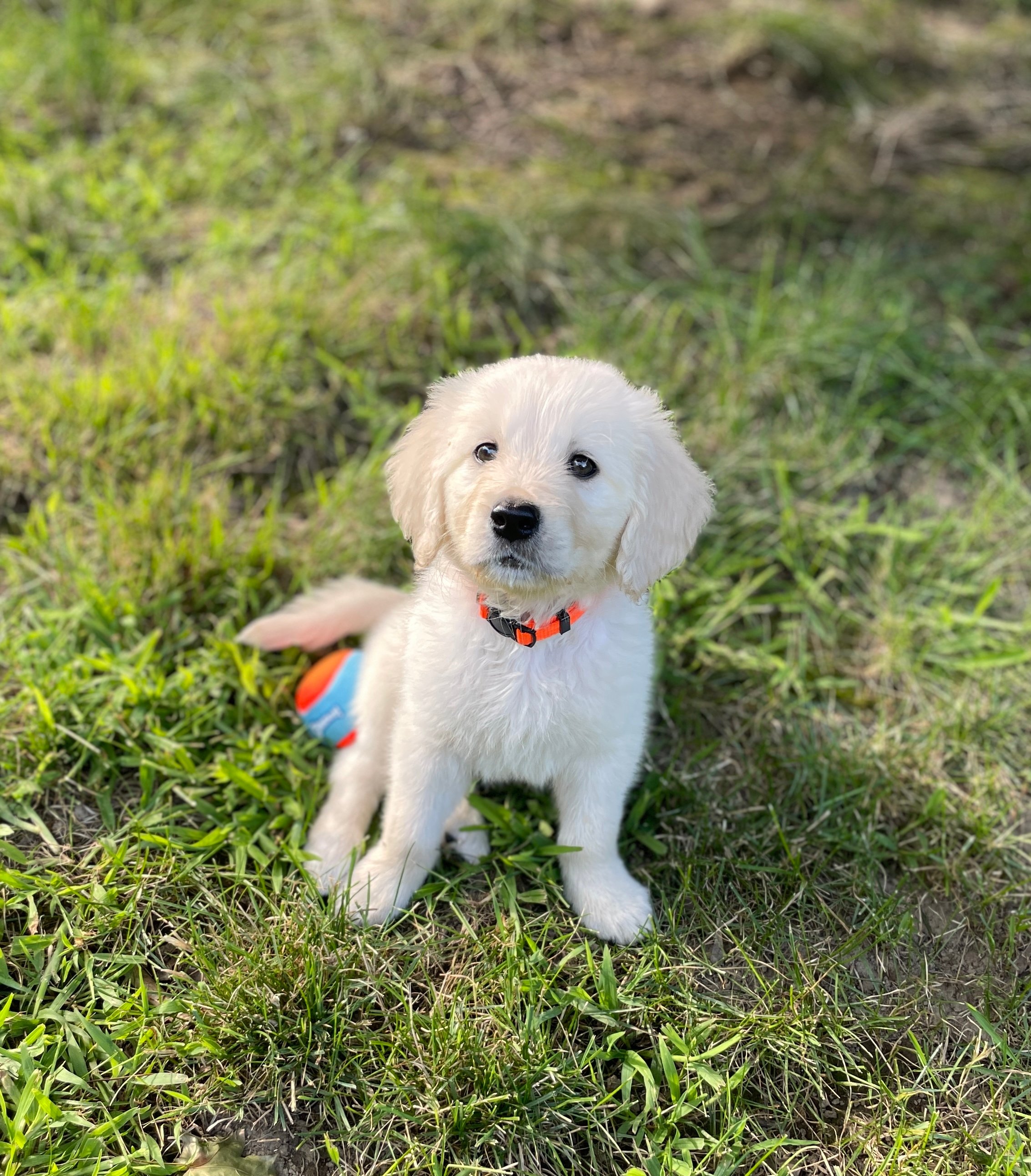
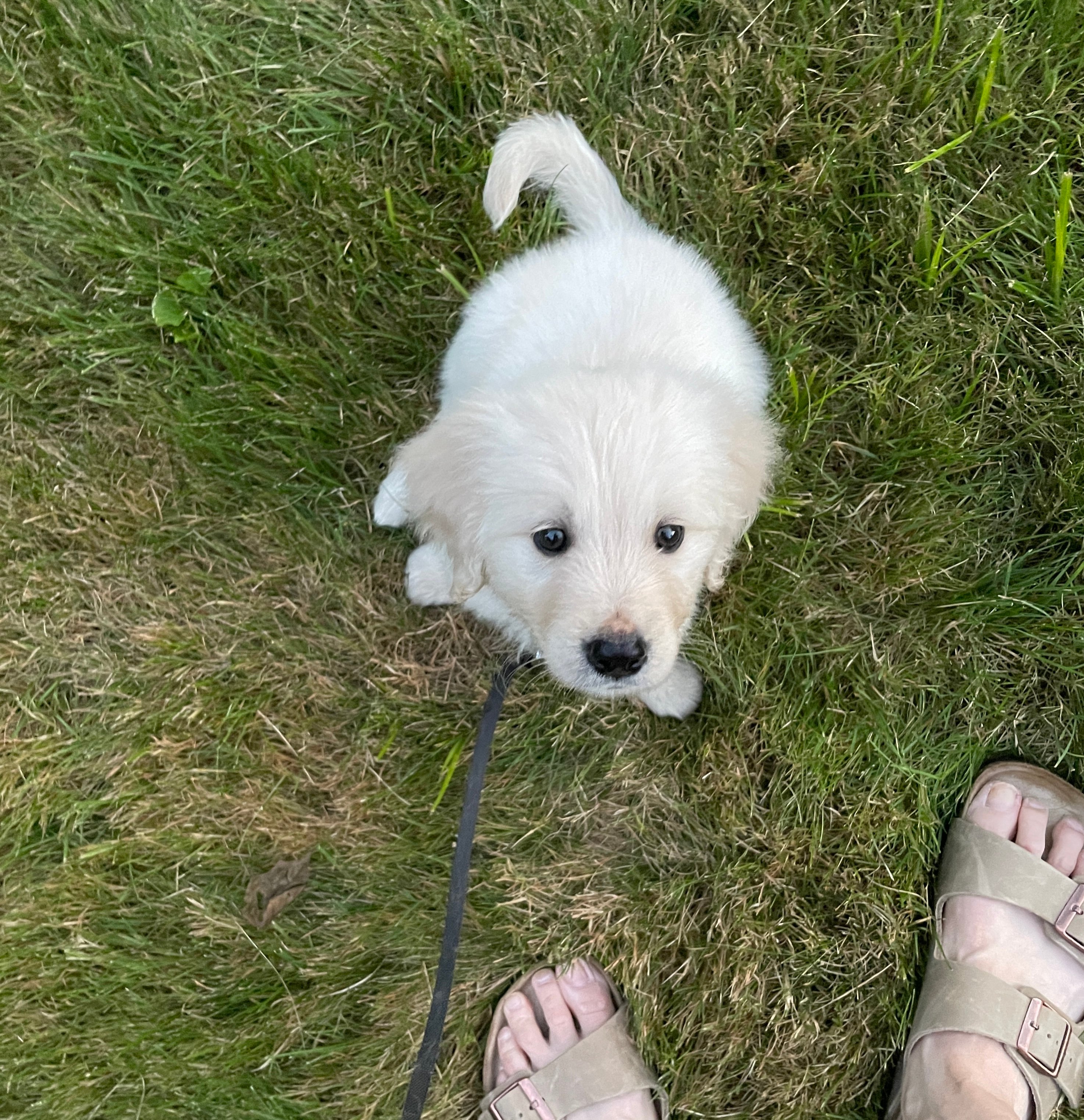
Shunt Hunt time for Rocket! 🚀 A "Shunt Hunt" is a clearly non-medical term for an ultrasound looking to rule in or out a PortoSystemic Shunt (PSS). It's challenging, but can be very rewarding.

To explain in less medical terms, a Shunt Hunt follows the normal "map" of blood vessels into and inside the liver. I am looking for a.... let's call it... Shunty Shortcut that the blood would take to skip the liver detoxifying step.
Like any good treasure hunt, there are helpful clues along the way. The history, bloodwork, clinical signs and other ultrasound findings before even looking at the liver.
And giving credit where credit is due - I learned everything I know about Shunt Hunts from Dr. Eric Lindquist at Sonopath. 💥
I brought my ultrasound machine to Dr. Barker's veterinary hospital (North Yarmouth Veterinary Hospital). Rocket had been dropped off at the hospital ahead of time, after being fasted for a conservative 6 hours - in retrospect, longer would have been better as Rocket is a hearty eater 😆. Food in the stomach can block needed views of the liver. We worked around it, but ideally an empty stomach is far easier.
Sedation is extremely helpful as well, to reduce the inevitable "puppy wiggles" while hunting for a shunt that may be mere millimeters in size.
With a team effort of restraint and a distracting puppy chew treat, we managed to complete the ultrasound and get the needed images. After some post-scanning consultation with other specialists and combing through the ultrasound clips image by image, we found it! Woo-hoo!
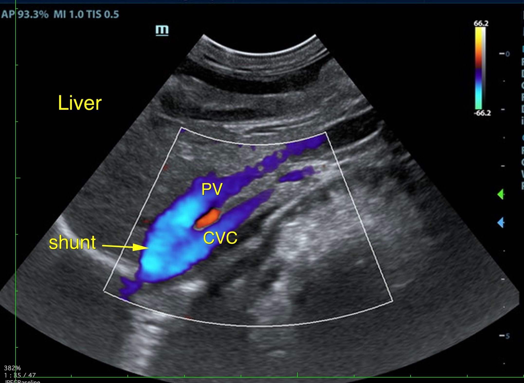
We suspected a PSS (ie Shunty Shortcut) deep in the liver up by the diaphragm. This is an Intra-Hepatic shunt. This particular kind, a Right Divisional, can be very short and small. Easy to miss.
Tidbit of information: Color doppler as shown above shows you directional flow, in this case blood flow.
The acronym BART - Blue Away, Red Toward, helps us remember what we are looking at.
Blue is blood moving away from the ultrasound probe (ie the Top of the screen). So the above picture shows blood from the Portal vein (blood heading to be detoxified by the liver - Normal) taking a Shunty Shortcut down into the CVC (caudal vena cava - blood headed to the heart to be pumped out to the body). Those two vessels shouldn't attach with blood flow in that direction. Confusing and tricky, right ??
So.... now what?
We strongly suspected the Intra-Hepatic Shunty Shortcut but ideally needed a CT to confirm and figure out what Rocket's treatment options are. Not all shunts can be fixed, and we need some pretty detailed information to make those decisions.
But this sweet little face...
A week after Rocket's ultrasound was done, I was planning a work trip to instruct and lecture at a veterinary ultrasound lab in Andover, NJ at Sonopath Education Center. Right next door to the Education Center is the Sonopath Imaging Center.... with a fancy new CT machine! I'd been dying to see the machine in action, and here's a puppy who NEEDED a CT. 🤔.
I reached out to Dr. Eric Lindquist and Dr. Ken Leal at Sonopath, and they generously offered to do the CT on Rocket for no charge if I brought him down with me. They will likely write a Case Study on Rocket when everything is done.
Dr. Barker gave me permission to bring Rocket to NJ with me, and authorized his needed care to get a CT done.
CT.... here we come!! How hard could it possibly be to bring a tiny 6 lb puppy down to NJ with me? 😂😂😂
Stay tuned for Part 3 to find out! 😄